Macular Pucker
Common Retinal Disorders
Macular pucker
The macula of the retina provides our clearest, central vision. A condition called macular pucker can affect this central vision, and occur from several sources. The macula has a high concentration of photoreceptors called cone cells. Macular pucker may occur from changes in the vitreous of our eyes or in those with diabetic retinopathy. It can also occur from eye inflammation or after eye surgery.
Certain conditions can cause a type of scar tissue to form on the surface of the retina. This tissue may form over the macular region as epiretinal membranes. They can contract and cause the retina underneath to fold and wrinkle. This can affect vision and may require surgery to repair.
Floaters and Flashes of Light

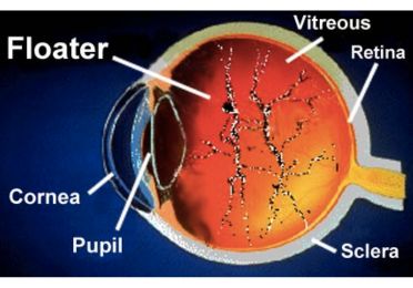
The normal eye is filled with a gel-like material called vitreous humor. On the back wall of the eye is the retina, which absorbs the light from the world around us. Over time, the vitreous can undergo change. Small collagen condensations called floaters may form. They can appear to move in our vision.

Floaters cast shadows onto the retina that we see. They may appear as webs or dust in our view. Most are harmlessly annoying.
Flashes of light, however, may be more serious. This may signal that vitreous is pulling on the retina.
Vitreous Detachment
The vitreous gel can liquefy and shrink with age. This can cause the vitreous to detach from the back wall. This is called a posterior vitreous detachment. It may appear as moving veils or clear curtains in our vision.
Retinal Tear
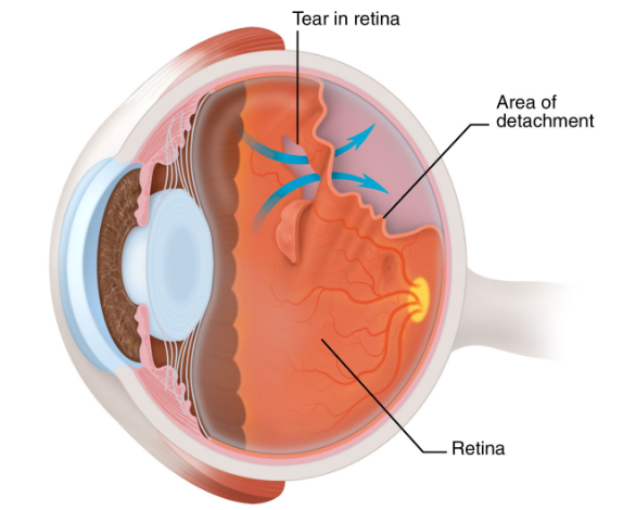
Sometimes, the vitreous can pull on the retina. This pulling is called vitreous traction. Traction and vitreous changes can eventually tear the retina. Laser may be used to treat such areas. This aims to prevent fluid from going behind the retina.
Retinal Detachment

Fluid may pass through a tear or hole in the retina. Fluid behind the retina detaches it from the back wall. Vision is lost in this area.
Retinal Detachment Surgery
Surgery may repair a retinal detachment. First, the fluid behind the retina is drained. Silicone exoplants are placed external to any tears or holes. An encircling band is placed on the outside of the eye. This repositions the retina back against the inner wall. Laser retinopexy seals the holes or tears in the retina. This aims to prevent fluid from passing through them. A gas bubble may be used to hold the retina during healing. The gas bubble reabsorbs and the surgery is complete.
To summarise, Retinal detachment is a very serious eye problem. It can occur without warning at any age, or occur from eye trauma. People who are nearsighted are at greater risk. Some signs of a potential retinal detachment are detectable by your eye care provider. Regular eye check-ups are very important in detecting these early signs. Any noticeable changes in your vision, especially flashes, or sudden changes in your floaters, should be examined by your eye care provider immediately. Although complex, modern microsurgery affords repair of retinal detachment, and may restore vision, depending on specific circumstances of each case.
Contact Us
If you have any other questions or concerns, please contact us at:
The Eye clinic
Dr Swati Agarwal -The Eye Clinic
2A Motilal Basak lane
Kakurgachi
Kolkata, WB 700054
www.swatiagarwal.in
+91 9147714355
[email protected]
Important Note
This information is provided for general educational concepts only. Your specific circumstances, conditions, treatment approaches, and results may vary from those shown here. Additional information, instructions, and precautions are to be provided by and discussed with your eye care provider.
Need Urgent Help?
Call Us: